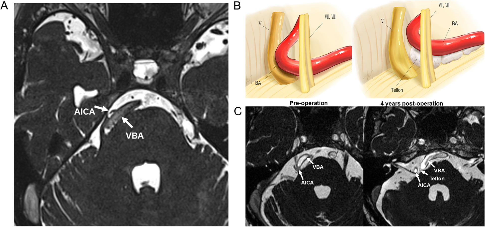
Fig. 1

An image of tortuous dolichoectatic vertebrobasilar artery (VBA) and AICA. a An image produced by a common 3D-TOF magnetic resonance imaging (MR) shows a tortuous dolichoectatic VBA and the AICA stem were shifted to the right side. The trigeminal nerve is curved and deformed due to compression of the translocated arteries. b A schematic diagram of the surgical procedure. Cigarette-like Teflon implants are placed between the VBA and the nerve for decompression. c Preoperative (pre-op) and 4 years postoperative (post-op, 4 years) MR images. In pre-op MR, the bilateral vertebrobasilar artery (VBA) is seen to curve towards the right side and compresses the trigeminal nerve as well as the 7th and 8th cranial nerves. The 4 years postoperative image shows that Teflon remains between the VBA and the brain stem without shifting isolated the trigeminal nerve and arteries, but it simultaneously compresses the 5th, 7th, and 8th cranial nerves, which is the cause for recurrence of facial pain