Fig. 3
From: Diagnosis of a subarachnoid hemorrhage with only mild symptoms using computed tomography in Japan

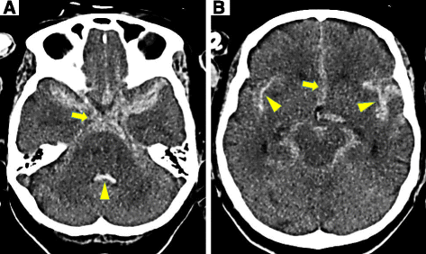
Typical non-contrast CT images of SAH showing blood in the subarachnoid space. The slice is at the level of the suprasellar cistern in a. Blood filling the basal cisterns, and there is hyperattenuating blood in the suprasellar cistern spreading laterally into the medial portion of the Sylvian fissures. In addition, there is blood that has refluxed into the fourth ventricle. An arrow indicates the blood in the suprasellar cistern. An arrowhead indicates blood in the fourth ventricle. The slice is at the level of the midbrain (mesencephalon) in b. There is blood in the perimesencephalic cistern extending anteriorly into the anterior interhemispheric fissure at the level of the midbrain. Blood extending bilaterally into the Sylvian fissures. An arrow indicates the blood in the anterior interhemispheric fissure and the blood in lateral extents of the Sylvian fissures are indicated by arrowheads